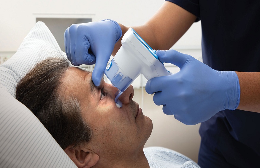
In the fascinating realm of neurological assessments, where the complexities of the brain are unraveled, the eyes stand as a remarkable gateway to understanding its intricate workings. Beyond being a routine check, the evaluation of pupillary reaction emerges as a pivotal and indispensable diagnostic tool. However, with the utilization of cutting-edge neurological instruments, this evaluation transcends its conventional limits, achieving an unprecedented level of precision.
The remarkable insights garnered through this refined process hold the potential to revolutionize patient care as healthcare professionals gain a deeper understanding and make well-informed decisions to improve the outcomes of their patients. By harnessing the power of advanced technologies, we unlock a new era in neurological assessment, where the eyes unveil invaluable secrets that help shape the future of compassionate and effective medical treatment.
Understanding Pupillary Reaction
At its core, pupillary reaction encompasses the intricate mechanism through which the pupil, serving as the gateway to our visual world, intricately responds to a myriad of stimuli, with an emphasis on the primary stimulus being light. This remarkable physiological process is a reliable gauge for assessing the complex neurological system’s delicate balance and optimal functioning. Hence, any discernible deviation in the size of the pupil or its inherent responsiveness acutely raises the proverbial red flag, potentially unraveling hidden neurological abnormalities that might otherwise remain clandestine, evading detection, and eluding timely intervention.
By manifesting as a telltale sign, such anomalies reverberate as an alarming call, urging medical professionals to delve deeper into the intricate labyrinth of the nervous system, unlocking the secrets veiled beneath its enigmatic facade, and illuminating the path towards comprehensive diagnosis, effective treatment, and ultimately, the preservation of neurological well-being.
Neurological Tools for Evaluating Pupillary Reactions
The world of neurological tools is vast and ever-evolving. For pupillary assessment, tools range from the basic flashlight, used for a swift check, to advanced devices like pupillometers and infrared pupillography. These tools, especially the latter, offer quantitative data, making the evaluation process objective and accurate.
Preparing for Pupillary Evaluation
Before diving into the assessment, preparation is key. Ensure the patient is comfortably positioned, ideally in a semi-reclined posture. The environment plays a role, too; a dimly lit, distraction-free room can make a world of difference, ensuring the pupils react naturally without external influences.
Assessing Pupillary Size and Shape
With the patient ready, observe the pupils’ size and shape. Are they equal? Is one larger than the other, a condition known as anisocoria? Or perhaps they’re constricted (miosis) or dilated (mydriasis)? Though seemingly simple, these observations can provide a wealth of information about the patient’s neurological health.
Evaluating Pupillary Response to Light
Using a flashlight or another light source, shine a beam into each eye, observing the pupillary reaction. A healthy, brisk response indicates good neurological function. On the other hand, a sluggish or absent response can be concerning, pointing towards potential issues that warrant further investigation.
Assessing Pupillary Reactions to Accommodation
Beyond the light response, there’s the reaction to accommodation. Ask the patient to focus on a near object, observing the pupils as they do. A normal response would be constriction of the pupils. Any deviation from this can be indicative of neurological anomalies.
Pupillary Response in Neurological Conditions
Pupillary reactions can offer insights into a plethora of neurological conditions. For instance, a dilated pupil that doesn’t respond to light might indicate a traumatic brain injury. Similarly, certain drug intoxications can lead to pinpoint pupils. The pupillary response can vary in stroke cases, offering clues about the affected brain region.
Potential Limitations and Considerations
While neurological tools like the NPi pupilometer and pupil measurement techniques are invaluable, they come with limitations. Medications, for instance, can influence pupillary reactions. The lighting conditions, if not optimal, can skew results. Moreover, cooperation is crucial; an anxious or uncooperative patient can make the evaluation challenging.
Conclusion:
Evaluating pupillary reaction is an art and science combined. This assessment becomes a cornerstone in neurological evaluations with the right neurological tools. As medical experts, staying abreast of the latest techniques and understanding the nuances of pupillary reactions can make a significant difference in patient care. Whether it’s a routine check or a detailed neuro exam, always remember: the eyes don’t just see; they speak volumes about our neurological health.


